How Does Diabetic Retinopathy Develop?
Diabetic retinopathy, which develops as a result of diabetes, is the most common cause of blindness between the ages of 20 and 65. If left untreated, the risk of blindness is 25 times higher than in people without diabetes. The main cause of the problems in the retina is damage to the retinal blood vessels due to diabetes. As a result of this damage, microaneurysms (small bulges in the vessels), hemorrhages, edema (fluid buildup) and exudates (deposits caused by leakage from the vessels) are seen in the retina in the early stage of the disease, while in more advanced stages the nerve layer is deprived of oxygen (ischemia) and new blood vessels and membranes form inside the eye.
In the final stage of the disease, blindness develops with more serious consequences such as bleeding into the eye from these new vessels and the membranes pulling the retina away from the wall of the eye (retinal detachment). For this reason, early diagnosis followed by appropriate monitoring and treatment is very important in these patients. With today's modern technology, the rate of blindness in these patients can be reduced to below 5%.
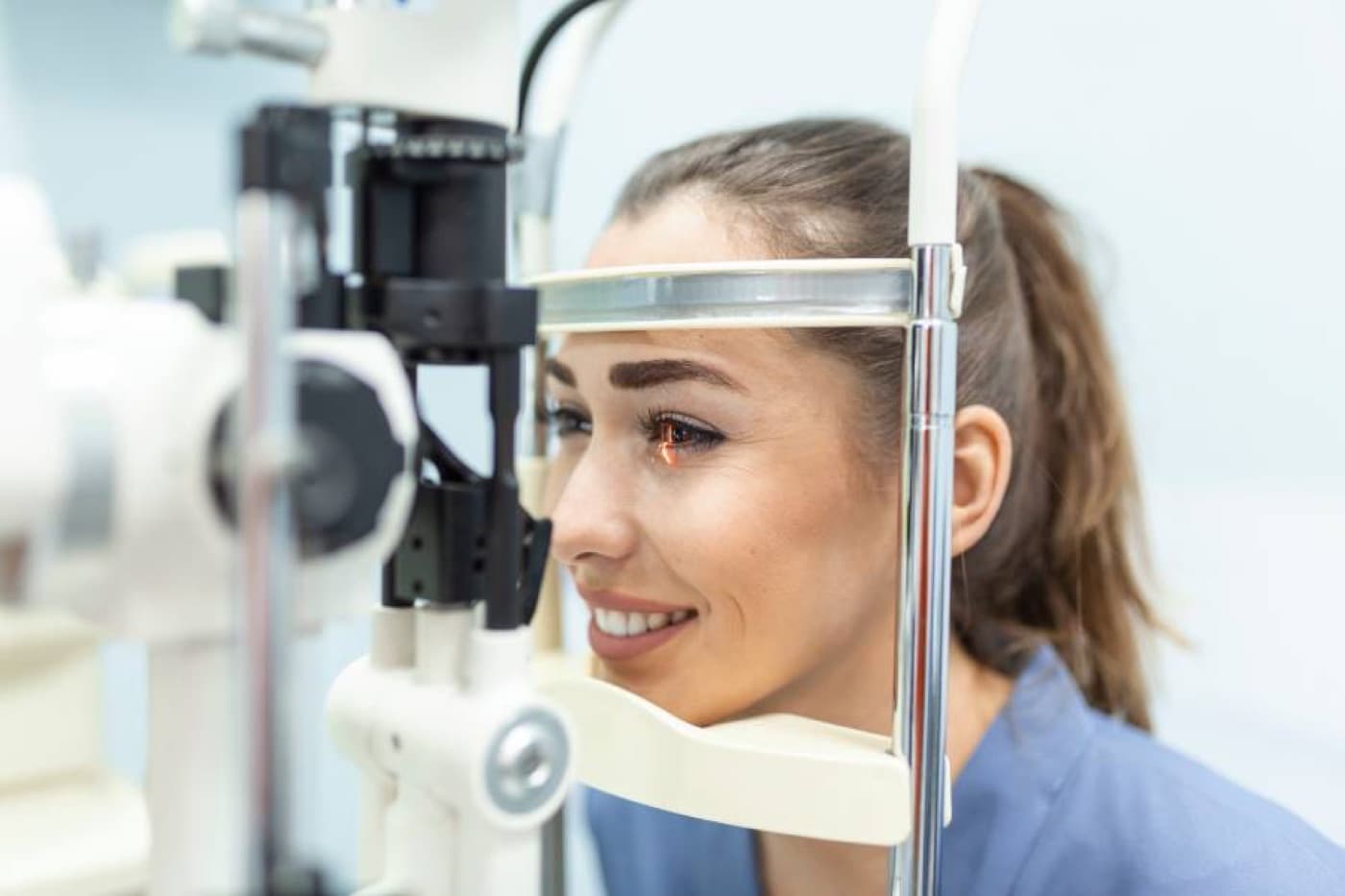
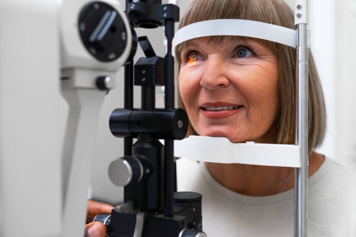
Patients with diabetes should be examined by an ophthalmologist as soon as they are diagnosed and should continue their check-ups as often as their doctor recommends, with no more than a year between visits. During these examinations, the ophthalmologist may perform an eye angiography when needed. This procedure, performed while the patient is seated after a dye is injected into a vein in the arm, takes a few minutes and allows a more detailed examination of issues such as the location of leakage, ischemia and the presence of new vessels in the retina. At Can Göz Kliniği, imaging can also be performed without dye using OCT angiography. Another examination method frequently used in this patient group is optical coherence tomography (OCT). With this method, the retina can be examined within seconds at a resolution of 5–10 microns.
The main goal in treating diabetic retinopathy is to preserve existing vision. The most commonly used treatment for this purpose is laser treatment. Laser treatment is a painless procedure. It aims to reduce edema in the retina and to make newly formed, bleeding-prone vessels inside the eye regress. At Can Göz Kliniği, treatment can be performed with state-of-the-art micropulse ("cold laser") technology without damaging the healthy nerve cells in the eye that provide vision. Because this laser does not harm the cells, it can be used repeatedly.
When laser treatment is not sufficient, medication can be given by injection into the eye. In advanced cases, surgery may be necessary. In a vitrectomy operation, which uses advanced technology and is performed by a surgeon experienced in this field, hemorrhages and membranes inside the eye are cleared and the detached retina is put back in place. At the end of the operation, gas or silicone oil may be placed inside the eye, depending on the situation.
The information on our website is not intended to direct individuals to any treatment. Do not take any diagnostic or treatment steps without consulting your doctor. The content does not contain information about İzmir Can Hastanesi's therapeutic healthcare services.